The focus of this post is an ammonite specimen with very complicated internal shell structure consisting of "sutures" or partitions. Please see the diagrams I provided in my previous blog on “Ammonite Sutures,” which I posted on Oct. 19, 2014.
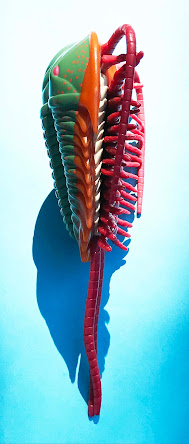
Ammonites are extinct mollusks (cephalopods) that range in geologic age from the Devonian to the end of the Cretaceous. The following image shows a replica of the general appearance of many ammonites and is not the same genus as the one discussed below. The outer shell of an ammonite conceals the internal shell structures (called septa), which can be much more complex than what is visible on the outside of the shell.
The fossil ammonite discussed below is moderately large (12.5 cm tall, 11 cm wide). The first and third images illustrated below show its sides, and the second image shows its narrow, flat venter edge (2.75 cm thick). The locality of this ammonite is in the South Dakota region. It was collected nearly a century ago by an anonymous collector. The outer shell is missing on this specimen, but its internal shell details are well preserved.
The interior of an ammonite shells have many chambers, and the shelly chamber walls are called sutures. The sutures constitute lines which have have loops in them. If a loop points toward the aperture (the large opening of the shell where the main part of the animal was located), the loop is called a saddle. If a loop points in the opposite direction, it is called a lobe. Taken as a whole, each chamber wall consists of saddles and lobes. The more complicated the suture (i.e., the more it has squiggles or frills, the younger the geologic age of the ammonite). In the case of the ammonite figured here, it can be readily seen that the suture pattern has a suture line with all of its loops frilled.
The suture pattern of the fossil ammonite shown here has very frilled sutures; so much so that they can be classified as ammonitic sutures. They help greatly in the identification of the family and the genus of an ammonite. Using its suture pattern, I was able to identify the family of this ammonite as Placenticeratidae and the genus as Placenticeras sp. Meek, 1876. This genus is Late Cretaceous in age and ranges in age from the late Cenomanian to the late middle Campanian. The genus is somewhat widespread in the world. In the United States, it has been found in the “Western Interior” region of Wyoming, South Dakota, Colorado, Utah, New Mexico, and Texas.
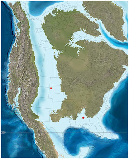
This paleogeographic map (http://jan.ucc.nau.edu) shows the distribution of the Western Interior Seaway (in light blue color), which existed during the time of Placenticeras.
It is important to mention that the outer shell of the above-illustrated specimen is mostly missing. In the identification of any specimen of Placenticeras it is critically important to know whether or not the outer shell had tubercles (where located on the shell and how many tubercles). On the specimen illustrate here, tThere are a few poorly preserved tubercules, but otherwise this information is not available. Identification of the species is, therefore, indeterminate. That is why it is always important to see as many specimens as possible when identifying a fossil.


