Friday, December 29, 2023

VERY MEMORABLE HAND-DRAWN SEASHELL ILLUSTRATIONS

Thomas Martyn (1760-1816) was an English zoologist interested in beetles and spiders. He was also a shell dealer who purchased seashells. In addition, he was a gifted, artist whose major work, The Universal Conchologist, was published in 1784 –1787 (Wikipedia, 2023). In his four-volume work, he and his team of apprentices, created numerous carefully done, hand-drawn and hand-colored portraits of many shells (some of which are rare) collected by Captain James Cook on his different voyages to the south Seas. Martyn later expanded his work to several volumes, each containing many colored plates. He wanted to have his work encompass “every known shell,” but he was not able to accomplish this “laborious, expensive, and arduous undertaking” (see Williams, 2015).

Martyn’s The Universal Conchologist is now worth a small fortune, starting at several thousands of dollars. Dance (1986, pp. 72–73) wrote that Martyn’s shell publications have a “beauty, seldom surpassed in the history of conchological iconography." But, because of the haphazard way Martyn named the species he illustrated, the Universal Conchologist is now considered a non-binominal [i.e., a non-scientific] work”] (Dance, 1986).

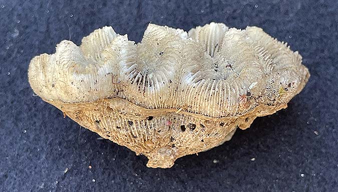
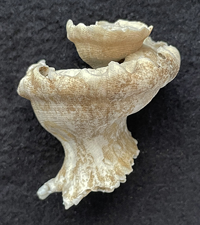
Two views (front and back) of Alcithoe arabica (Gemlin, 1791), family Volutidae. This gastropod, which is endemic to New Zealand, was originally referred to as the “Arabic volute,” because its zig-zag markings were thought to resemble Arabic writing. The species belongs to family Volutidae. Its shells, which can be up to 9 inches long, have color and variable markings. This species is subtidal, lives on soft sediments, and feeds on bivalves. In Martyn’s book, this species was shown as “figure 52w.”


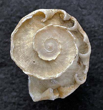
Two views (oblique top and bottom) of Astraea heliotropicum (Martyn, 1784). This deep-water marine gastropod, which is also endemic to New Zealand, was originally referred to by Martyn as the “sunburst star turban” or as the “circular saw shell.” This species belongs to family Turbinidae. The color of its shell is highly variable, and can be from white, gray, reddish brown, or blackish brown. This species lives in deep water. 


References Cited:

Dance, S. P. 1986. A history of shell collecting. E.J. Brill–Dr. W. Backhuys, Leiden, 1986. 265 pp., 31 pls.


Glaac.uk/myglasgow/library/files/special/exhibits/month/July2009.html


Wikipedia. 2023.


Williams, N. 2015. This laborious, expensive, and arduous undertaking. Thomas Martyn’s The Universal Conchologist. The National Library of Australia Magazine (June issue), pp. 16–17. 


Thursday, December 28, 2023

MINUTE SHELLS

Small-sized shells, which are also called "minute shells" or “micromollusks,” are not usually subjects in sea-shell books. Yet, these small shells are an important part of mollusk studies. They are usually collected as “grunge,” taken into the lab, and carefully sorted into different species. It can be pains-taking work, and most shell collectors would rather leave this kind of work to other enthusiasts. That is where a person like Bertram (Bert) Draper filled an important role. Bert liked to collect and sort “grunge.” Then he took it to another whole level, when he painstakingly glued numerous examples of common micromollusks (mostly gastropods, some bivalves, and a few scaphopods) onto a piece of sturdy paper and placed it into a single plastic box, with a closable lid (see image below). Each shell was also labelled as to its identification. He then donated these boxes to various shell organizations that annually held auctions to raise money for their yearly activities; especially awarding small-research grants to students doing their research on mollusks.


These plastic boxes became a familiar sight to Southern California shell collectors who attended annual shell meetings (with auctions) in the 1980s and 1990s. At one of these events, I bid on and bought one of these plastic boxes (shown above) filled with micromollusks. For me, it represents a treasurable acquisition because when Bert passed in 2000, the very useful plastic boxes with many minute shells (all properly classified) were no longer readily available at the auctions.


Within each plastic box, Bert also included a typed list of all the minute shells contained within, as shown in the above image.


Close-ups of two of the examples of the minute mollusks that Bert put into each of his plastic boxes.


I knew Bert and liked the man. Whenever I visited the Malacology Collections at the Natural History Museum of Los Angeles County, Bert was usually around because he was a volunteer who helped in micro-photography. He was an accomplished expert in micro-photography of seashells, and he gave me some good tips on how to photograph my fossil examples. For information about him, I encourage you to read the article by Groves and McLean (2001).


In the years between 1972 and 1974, Bert wrote several articles (at least 9) for The Tabulata, a now discontinued newsletter concerning the study of seashells (malacology). These articles concern various minute-shell species and also how to photograph minute shells. An article written by him (Draper, 1972) is especially relevant to this blog post. 


References Cited:

Draper, B. 1972. Minute shells-part 1. The Tabulata, 5(4), 3–6. https://www.scamit.org.tools >Mollusca 


Groves, L.T. and J.H. McLean. 2001. Bertram C. Draper 1904–2000. The Festivus 6:63–65. This publication can be accessed via biodiversitylibrary.org and also via

https://researsch.nhm.org> dataimages > Draper


Saturday, December 23, 2023

WHEN IS A S0-CALLED FOSSIL REEFTRULY A REEF?

The answer to that question is: when field evidence shows unequivocally that any so-called reef” can be proven to have been constructed by wave-resistant/frame-building organisms, and these remains are in situ at their site of origin.


In the published literature, however, there are some so-called reefs,” when in reality, they are actually pseudoreefs. Their sedimentary bedding planes by been masked by weathering, thereby creating massively looking structures that can resemble non-bedded true reefs. Even more critically important, a reef fauna is not present.


A case in point is discussed here. It was the topic of my masters degree in geology (Squires, 1968), which focused on Permian marine rocks in Last Chance Canyon, southeastern New Mexico. Based on spending a summer of studying several so-called “reefs”, I found no evidence that could be used to paleontologically or lithologically differentiate these rocks from the surrounding, laterally equivalent strata. The only fossil material present consisted of broken fusulinids, fragments of brachiopods, and occasional crinoid columnals. These fossils together usually comprised no more than 10 percent of the rock in question, which consisted of dolomitized mudstones and muddy wackestones.


Pseudoreef (area "2," outlined in dashed lines) in upper part of upper member of the San Andreas Formation (from Squires, 1968).


Pseudoreef (outline in dashed lines = area between 1 and 2) in upper part of upper member of the San Andreas Formation (plate 10 from Squires, 1968).


Photomicrograph of a typical example of the dolomite rock that comprises the pseudoreefs (plate 3 from Squires, 1968). 


Although the pseudoreefs do have massive-looking outcrops that resemble true reefs, this resemblance is only superficial and is related to weathering associated with steep-walled, south or south-facing outcrops. Another complicating factor is that eastwardly, within the studied area, the San Andres Formation interfingers with the sandstone tongue of the underlying Cherry Canyon Formation, which locally can have moderately large channel-and-fill structures. This interfingering relationship makes it seem like the eastward sides of the pseudoreefs transition into reef-talus debris,” but this is not the actual case because these supposedly reef-talus beds” consist of sandy dolomite with some fossils, but with no reef-forming fossils.


My pseudoreef interpretations are also supported by the work (unknown to me at the time) of Harrison and Jacka (1967), who reported that in the area of Last Chance Canyon, the San Andres Formation and the Cherry Canyon sandstone tongue accumulated in a deep-water submarine canyon. The fossils in these two units are death assemblages of shallow-marine species (e.g., fusulines, bryozoans, brachiopods, crinoid columnals, and echinoid spines) that were transported by submarine currents into deeper waters (approximately 600 to 1,000 feet deep).


References Cited: 


Harrison, S.C. and A.D. Jacka. 1967. Depositional environment of Cherry Canyon Sandstone Tongue, Last Chance Canyon, New Mexico. American Association of Petroleum Geologists Bulletin 51(3):p. 268 (abstract). Only the abstract is free.


Squires, R.L. 1968. Origin of reeflike masses in the upper member of the San Andres Formation, central Guadalupe Mountains, Eddy County, New Mexico. Master of Science Thesis, University of New Mexico, 124 pp.


Saturday, December 16, 2023

THE CHALLENGE OF IDENTIFYING EXTANT COLONIAL-STONY CORALS


Colonial-stony corals (also called hexacorals or scleractinians) belong to phylum Coelenterata. The geologic range of colonial-stony corals is Middle Triassic to modern day. This blog post concerns the hematypic hexacorals, which are the ones that harbor symbiotic algae and, therefore, can be reef builders. 


A reef is a wave-resistant, ridgelike or moundlike structure, built from the remains of reef-building organisms (colonial corals, red algae [e.g., Lithothamnion], green algae, as well as the remains of mollusks, echinoderms, crustaceans, bryozoans, microscopic foraminiferans, and other animals). A reef also consists of in situ organic-constructed unstratified rock. If a wooden ship hits a reef, it could sink the ship! Only the upper crust of a reef is alive and growing. Coral reefs have always been are restricted to the tropics in shallow-warm waters. Today, coral reefs occur between latitudes 30°N and 30°S. 


One might think that the identification of reef corals is simple, but the process is not easy. There are at least 100 genera of modern-day, reef corals, but there are only a few (less than 10) basic shapes (e.g., massive, branched, columnar, domed, sheet-like, furrow-like). And these shapes are not confined to any one genus (Wood, 1983). The skeletons of colonial corals are referred to as the corallum, and the individual polyps (corallites) that comprise these skeletons can be very similar looking. These similarities include the size, spacing, and number of septa (radial partitions) in the centers of the individual polyps. That is why the title of this blog post is appropriate.


             note: In the figures shown below, each respective

                         caption is just below each figure.                           

Styloceniella? sp. (two views: overall coral corallum and a closeup). The specimen is 14 cm tall and 10 cm wide. It has an overall “domed” shape with small knobs. The polyps are crowded closely together, and the septa within the polyps are short and small. According to Wood (1983, p. 71), this genus is confined to the Indo-Pacific region and often grows in crevices or on the undersides of rocks or other corals.


Echinophora sp. (two views: oblique side and top). The specimen is height 6 cm and width 7.5 cm. The genus is found in the Indo-Pacific region. Echinophora is a “shape shifter” as its skeleton can be from branching, flat, fan-like or leaf like, or even the very distinctive wide-cup shape (like the specimen illustrated here).


Fungia sp. (two views: top and side). The specimen is 3.4 cm wide and 0.5 cm thick. This distinctive Indo-Pacific genus is commonly referred to as the “mushroom coral.” Juveniles are often found in crevices or other sheltered places. Small-sized Fungia are capable of movement by using their tentacles at the edge of their shells. Rubble-strews slopes are often densely populated with fungiids. There are probably 15 to 20 species of Fungia (Wood, 1983, pp. 117–118).


Oulophyllia? sp. (three views: side, top, and closeup or a single corallite). The specimen is 6.7 cm long, 4 cm wide, and 3.5 cm tall. It has a meandroid shape, with “valleys.” It is an Indo-Pacific coral.


Pavona sp. (Three views: coral head, close up, and more close up). This heavy specimen is 28 cm long and 21 cm wide. It is an Indo-Pacific coral and is fairly common. There is great variability in morphology of this coral genus (Wood, 1983, p. 90). 


Sideastrea sp. (three views: coral head, close-up, and more close-up).  The specimen is attached to the side of a large (30 cm long and 28 cm wide) gastropod shell of Strombus gigas. This coral is an Atlantic reef species that lives in the Caribbean area (e.g., Bermuda It is a fairly uncommon coral that appears to prefer reef slopes). 

Reference Cited:


Wood, E.M. 1983. Corals of the world. T.F.H. Publications. 256 pp. 


Tuesday, December 12, 2023

CORAL-SNAIL EXAMPLES

Subfamily Coralliophilinae is a large group (200 to 250 species) of warm-water marine gastropods that feed exclusively on anthozoans (especially corals); hence, they are referred to as "coral snails." These gastropods burrow into corals or live in rock crevices, as well as on rocks or on sea fans, in shallow to deep waters (Eisenberg, 1981, p. 188). They are carnivores and feed on soft corals, hydrozoans, and anemones. The embryonic forms of coralliophilines allow for wide dispersal of their spawn; especially in the southwest Pacific (e.g., Japan, Taiwan, Phillipines, etc.). Coralliphilines are commonly  characterized by rather medium-size shells with some or numerous delicate spines. Some of the species show considerable morphologic variation, thus they have been very prone to being overnamed by workers.


Early workers assigned many of the coralliphilines to genus Latiaxis. A few of these gastropods do actually belong to that genus, but, in recent years, modern workers have reassigned many of these gastropods to various other genera. Three species of coralliphilines are illustrated below:


Latiaxis pilsbryi Y. Hirase, 1908. This name is still valid today, according to WoRMS (2023). Three views: front, back, and top, respectively. Specimen 35 mm (width) and 27 mm (height). This species is characterized by a flat spire rimmed by blunt spines. This species, which has variable morphology, can be found in the waters off Japan and Taiwan.
_______________


 Latiaxis mawae (Gray, J.E. in Griffith and Pidgeon, 1834). Four views: front, back, top, and right side, respectively. Specimen is 40 mm (width) and 52 mm (height). The aperture of this specimen has its operculum present. This species can be to 70 mm in height, and some specimens have a reddish brown color (see Hardy’s Internet Guide to Marine Gastropods). This species, which has variable morphology, can be found in the waters off Japan and and Queensland, Australia. The species mawae is the type species of genus Latiaxis (i.e., it is the species used to originally define the genus Latiaxis).


___________________


Babelomurex spinosus (Y. Hirase, 1908). Three views: front, back, and left side, respectively. Specimen: width 27 mm (width) and a 30 mm (height). This species has variable morphology and can closely resemble several other species. All? (most?) of them probably belong to a “species complex” that needs DNA studies. This species can be found in the waters off of Japan, Philippines, the southern China Sea, and North Australia. I used  WoRMS (2023) to help me with the vey confusing taxonomic history of this gastropod, which has many synonyms (previous names); and until [recently, including Latiaxis pagodus (A. Adams, 1853). 


References Cited

Eisenberg, J.M. 1981. A collector’s guide to seashells of the world. McGraw-Hill Book Company, New York, 239 pp.


Hardy’s Internet Guide to Marine Gastropods. An internet source with illustrations of museum-quality seashells.


Hirase, Y. 1908. On Japanese marine Mollusca 2, with the descriptions of two new species of Muricidae and Buccinidae. The Conchological Magazine 2:66–74. Available online.


WoRMS. 2023. An internet source.


Sunday, December 10, 2023

THE CARRARA MARBLE LOCALE IN ITALY

Carrara is a coastal region adjacent to a mining district, of the same name, in northwest Italy. The quarries are in the northern-most tip of the Tuscany region, at the northern end of the Apennines Mountains, which form the northwest-southeast trending backbone of the county of Italy. The Apennines intersect the east-west trending Alps, which occur along the northern boundary of Italy. The quarries in the vicinity of the town of Carrara have yielded more marble than any other place on Earth. 


A successive- closeup series of three “Google Earth images” showing the Carrara Marble-quarries area [denoted by the red arrows in the first two images]. 

Marble is a metamorphic rock that forms by heat and pressure applied to the sedimentary rock limestone. The original limestone in the Carrara area was deposited in an epicontinental-platform setting, during the early Jurassic time (almost 200 million years ago). Much later, in Tertiary times, polyphasic tectonic/metamorphic deformation took place and resulted in the fossiliferous limestone being metamorphosed into marble.


The Carrara Marble has been quarried (mined) since the time of Ancient Rome, over 2,000 years (22 centuries ago). Today, there are over 650 marble sites (about half are now abandoned) in the mountain area just east of the town of La Spezia that have extracted this rock from the Earth. Collectively, as shown above, the quarried area is readily recognizable on satellite images. The extensive man-made exposures of the white rock in this area appear as perpetual snow.


The Carrara Marble is world-famous because it is a “statuary marble,” which means it has the texture and durability that sculptors (including the Renaissance sculptor Michelangelo) want(ed) when creating their statues. Statuary marbles are generally whiter with fewer veins, thereby having a more uniform appearance that most marbles, and they are compact with a high quantity of calcium carbonate and a low quantity of silica.


Carrara Marble can be, as mentioned above, 1) pure white (statuario grade—which had a dedicated use for monumental sculpture because it has high tensile strength, can take a super-high gloss and polish, and can hold very fine detail; its supply now depleted since the late 1900’s), or 2) with an attractive, gray veining. Surprisingly, Carrara Marble is affordable (about $2 [in USA currency] per square foot). It is even sold at many home-improvement stores. Also, one should not use acidic liquids nor soap on this marble because they can cause the stone to darken. Carrara Marble can be used for countertops, but it requires proper sealing.


A representative cut and polished slab of Carrara Marble (1 square foot).


Recommended Online Reading/Viewing:


Bonne, K. 2020. Carrara Marble: from the sea to Michelangelo’s workship. Gondwanatalks.com/l/carrara-marble


Stein, C. and E. Sciolino. 2023. State of the Art. Smithsonian Magazine (December issue), pp. 31–41.


Wikipedia, 2023.


Friday, December 8, 2023

OFFICIALLY DESIGNATED STATE FOSSILS

Recently, while working up my post on the state fossil of California (see my Dec. 3, 2023 post on the saber-tooth Smilodon fatalis), I became curious as to what are the other state fossils of the United States.


I used the following excellent website (which lists the fossil names and also provides an illustration of each fossil:


 en.wikipedia.org/wliki/List_of_U.S._state_fossils 


I discovered that most states have an official state fossil, but only four do not: Arkansas, Hawaii, New Jersey, and Rhode Island.


Three other states: Iowa, Minnesota, and New Hampshire were formerly holdouts, but recently they tentatively proposed state fossils.


Most of the state fossils were designated as such in the 1980’s, and most of these state fossils are vertebrates (dinosaurs or mammoths).


A few states designated fossil mollusks as their state fossils; these are:


Maryland: shallow-marine gastropod Ecphora garderae of Miocene age.


Tennessee: shallow-marine bivalve Pterotrigonia thoracic of Cretaceous age.


Virginia: shallow-marine bivalve (pectinid = scallop) Chesapecten jeffersonius of Cenozoic age.


Furthermore I discovered that elsewhere in North America, only Nova Scotia in Canada has an official fossil: Hylonomus lyelli, a lizard-like reptile of Carboniferous age .

Sunday, December 3, 2023

SMILODON FATALIS: CALIFORNIA’S STATE FOSSIL

Smilodon fatalis is well know to most people interested in late Pleistocene Ice-Age fossils, especially if they have visited the Rancho La Brea Tar Pits and Page Museum in Los Angeles. The word “Smilodon” means “scalpel” and is derived from: “two edged knife” combined with tooth.


Smilodon fatalis is the second most common (dire wolves are the first most common) large fossil found in the La Brea Tar (asphalt) pits in Los Angeles, California. Over 100,000 bones of S. fatalis have been recovered from these pits.


Figure 1. Plastic model of S. fatalis.


Figure 2.  Smilodon fatalis skeleton, Page Museum, Los Angeles, Los Angeles County, California. 


Smilodon fatalis was a true sabretooth cat, as opposed to the sabre-tooth marpusial carnivore, known as Thylacosmilus, which lived only in South America during the late Miocene and Pliocene. 


Sabertooth cats belong to the cat family Felidae. There are two subfamilies in this family: the extinct Machairodontinae (sabertoothed cats) and the extant Felinae (true cats).


In size and weight, S. fatalis was as large as a modern-day African lion, but unlike modern-day large predatory cats, S. fatalis was a stealth, ambush predator with strong front legs and relatively light hind legs. Thus, it was not a swift runner. Smilodon fatalis has only 26 teeth, fewer teeth than in other cats. Additionally, the sharp saber teeth (upper canines) of S. fatalis were most likely used stab into skin/flesh and/or bite open the soft underbelly of its prey, rather than biting into bones in order to subdue its prey (as in large cats living today). Also, the enlarged, but relatively narrow upper canines of S. fatalis have a distinct backward curve in order to minimize resistance and they have tiny serrations on the inside of the curve.


Figure 3. Smilodon fatalis skull.


Figure 4. Another Smilodon fatalis skull.


Figure 5. Smilodon fatalis canine tooth; 9 inches (23 cm) long from “tip to bottom of tooth.” Widest part of tooth is 1.5 inches (4 cm).


Figure 6. Closeup of previous image of S. fatalis canine tooth. Notice the presence of tiny serrations on the inside edge of this canine. 


Figure 7. Smilodon fatalis jaw muscles (exhibit at Page Museum).


Figure 8. Smilodon fatalis skulls showing progressive replacement of canine teeth (exhibit at Page Museum).


Smilodon is known only from Cenozoic deposits in North and South America. There are three known species. They are from oldest to youngest:  S. gracilis [2.5 mya to 500,000 years ago] and known only with certainty from North America; S. fatalis [1.6 mya to 10,000 years ago] and known only with certainty from North America (California, Texas, Mexico, Nebraska, and Florida]; and S. populator [1 mya to 10,000 years ago] and known only from South America (Wikipedia, 2023). The occurrence of the land bridge (GABI = Great American Land Bridge, which enabled exchange of species between North and South America during the late Pliocene [see one of my earlier posts on GABI], was responsible for the dispersal of Smilodon from North America to South America. Smilodon populator  Berta (1985) did detailed research on the South American species Smilodon populator.


Figure 9. Smilodon gracilis. This skull, which is/was on display at the San Diego Museum of Natural History, is most likely from Southern California (Anza Borrego Desert region). According to Shaw and Cox (1986), fossils of this sabertooth cat have also been found in Florida
and Pennsylvania. 

References Used:


Berta, A. 1985. The status of Smilodon in North and South America. Contributions in Science, no. 370, 14 pp. [free pdf]


Lindsey, E.L. (ed.). 2018. Rancho La Brea: Treasures of the tar pits. A Natural History Museum of Los Angeles County publication. Third edition. 72 pp.


Savage, R.J.G. and M.R. Long. 1986. Mammal evolution, an illustrated guide. British Museum (Natural History). 258 pp.


Shaw, C.A. 2001. The sabertoothed cats. In, Rancho La Brea: Death Trap and Treasure Trove, pp. 26–27. Terra. Natural History Museum Special Edition, v. 38, no. 2.


Shaw, C.A. and S. Cox. 2006. The large carnivorans: Wolves, Bears, and Big Cats. In Fossil Treasures of the Anza-Borrego Desert. The last seven million years. Sunbelt Publications, San Diego, California. 394 pp.


Stock, C, (revised by J.H. Harris). 1992. Rancho La Brea, a record of Pleistocene life in California. Seventh edition. Natural History Museum of Los Angeles County, Science Series 37, 113 pp.


en. Wikipedia. 2023. Smilodon.